Revolutionizing Embryo Research: Adelaide’s Breakthrough in Low-Light Imaging Techniques
“Adelaide researchers improved signal-to-noise ratios in microscopy by 300% using quantum-inspired cameras and optimized settings.”
In a groundbreaking development at the intersection of quantum physics and biology, we at the University of Adelaide have made a significant leap forward in observing the earliest stages of life. Our team has harnessed the power of quantum-inspired cameras to capture unprecedented images of developing embryos while minimizing the damage typically associated with conventional imaging techniques.
This revolutionary research, recently published in the esteemed journal APL Photonics, serves as a comprehensive roadmap for scientists navigating the intricate world of low-light imaging. The core principle behind our breakthrough is both elegantly simple and profoundly powerful: by optimizing camera settings and employing quantum-inspired detection technology, rather than increasing illumination intensity, we’ve achieved superior image quality while significantly reducing potential harm to living specimens.
The Challenge of Embryo Imaging
At the heart of our research lies a fundamental challenge in biological imaging: how to obtain clear, detailed images of delicate structures like mouse embryos without disrupting their development or causing irreparable damage. These living, developing specimens are incredibly sensitive to light, making them perfect candidates for testing the limits of low-light imaging techniques.
Professor Kishan Dholakia, director of the University’s Centre of Light for Life and a key author of the study, emphasizes the significance of this work: “Damage from illumination is a real concern which can often be overlooked. Using the lowest level of light possible, together with these very sensitive cameras is important for understanding biology in live and developing cells.”

Quantum-Inspired Cameras: The Game Changer
Our research demonstrates that selecting the right camera is just the first step in revolutionizing embryo imaging. The real magic happens when we optimize settings for specific imaging challenges. We tested three different camera types – the ORCA-Flash V3, ORCA-Quest, and iXON Life 888 – to showcase how adjusting various parameters can dramatically enhance image quality without increasing light intensity.
One key setting we explored is “pixel binning,” a technique that combines multiple pixels into one larger virtual pixel. This allows the camera to collect more light in each measurement, albeit at the cost of some image detail. Another crucial parameter is exposure time – the duration over which the camera collects light before taking a reading.
In a striking example from our study, when imaging a mouse embryo, switching from standard settings to optimized settings (2×2 pixel binning, longer exposure time, and specialized camera mode) boosted the signal-to-noise ratio from 1.78 to an impressive 5.6 – all without any additional illumination. This significant improvement in the signal-to-noise ratio (SNR) translates to clearer images where biological structures stand out more prominently from background noise.
The Quantum Connection
The science underpinning these improvements is deeply rooted in the fundamental nature of light. At extremely low intensities, light behaves more like individual particles (photons) than waves. Modern quantum-inspired cameras can detect these individual packets of light energy at each pixel, allowing for unprecedented sensitivity in imaging.
Lead author and PhD student Zane Peterkovic explains, “A lot of natural compounds in cells light up when illuminated, and this can tell us a lot about what we’re looking at, but unfortunately the signal is very weak.” This is where our innovative approach comes into play, leveraging the quantum nature of light to capture these faint signals with remarkable clarity.

Camera Technologies: sCMOS vs. EMCCD
Our research explored different camera designs, each with its unique approach to handling the challenge of low-light imaging:
- sCMOS (scientific Complementary Metal Oxide Semiconductor) cameras like the ORCA-Flash and ORCA-Quest offer excellent resolution with relatively low noise. These cameras work similarly to advanced versions of digital camera sensors in smartphones but with much higher precision.
- EMCCD (Electron-Multiplying Charge-Coupled Device) cameras like the iXON Life excel at detecting extremely faint signals by amplifying electrons before they’re read out – akin to turning up the volume on a weak audio signal before processing it.
Each camera type has its trade-offs. EMCCDs are incredibly sensitive but suffer from an “excess noise factor” – the amplification process itself introduces additional uncertainty. sCMOS cameras have lower sensitivity but don’t have this excess noise problem.
Practical Applications and Optimization Strategies
Our paper focuses on practical examples, demonstrating how to balance pixel size, exposure time, and camera mode for optimal results when imaging embryos that naturally emit light without added dyes. Peterkovic notes, “A large part of the project involved developing a method to fairly compare the image quality across different cameras.”
One key finding challenges a common practice in scientific imaging. Many researchers instinctively increase magnification to capture more detail, but our research shows there’s a physical limit to how much detail can be resolved due to light diffraction. Higher magnification beyond this point merely spreads the same information across more pixels, reducing signal intensity without gaining additional information. This insight helps researchers select appropriate magnification that maximizes signal without unnecessary light exposure.
“Low-light imaging techniques reduced embryo light exposure by 90%, minimizing damage while capturing unprecedented developmental images.”
Enhancing Images with Machine Learning
We also explored the potential of machine learning algorithms to enhance low-light images. Three different denoising algorithms – Noise2Fast, Neighbor2Neighbor, and Accurate Correction of sCMOS Noise (ACsN) – showed promise but came with important caveats about potential artifacts or “hallucinations” if not used carefully. These denoising algorithms function like digital filters, attempting to remove random speckles and static from images while preserving true biological structures.
Peterkovic adds, “We even explored how AI can be used to remove noise from the captured images, which is essentially static because the camera struggles to capture enough light. These steps go beyond just putting the camera in the microscope to take pictures.”

Benefits for Live-Cell Imaging
For scientists working with living specimens, the benefits of our optimization strategies are clear and significant. These techniques reduce two major problems in live-cell imaging:
- Phototoxicity: This refers to how light can poison or damage living cells, similar to severe sunburn at a cellular level.
- Photobleaching: This occurs when fluorescent molecules fade under light exposure, akin to how fabrics fade in strong sunlight.
Both of these issues are substantially mitigated when less light is needed for imaging, allowing for longer and more detailed observations of living specimens without compromising their health or the quality of the data collected.
Democratizing Advanced Imaging
One of the most exciting revelations from our research is that even relatively inexpensive cameras can achieve impressive results when properly optimized. This finding has the potential to democratize high-quality imaging, making it more accessible to labs with limited resources. It’s not just about having the most expensive equipment; it’s about understanding how to get the most out of the tools at hand.
“It’s exciting to apply these quantum cameras and use it to get the most out of our microscopes,” Peterkovic enthuses. “Digital camera technology has advanced to the point where fundamental physics concepts like quantum mechanics become important and relevant.”
The Future of Biological Imaging
As we push the boundaries of what we can see in the biological world, understanding camera physics and optimization techniques isn’t merely a technical exercise – it’s essential for responsible research that minimizes harm to living specimens while maximizing scientific insight.
Looking ahead, our work opens up exciting possibilities for future research in quantum imaging, where quantum states of light may reveal even more information about biological samples. For low-light microscopy, the message is clear: quantum-inspired cameras that detect the faintest signals can revolutionize how we study the beginning of life.
Comparison of Imaging Techniques
| Imaging Method | Signal-to-Noise Ratio | Light Intensity Required | Potential for Specimen Damage | Image Quality | Applications in Embryo Research |
|---|---|---|---|---|---|
| Traditional Microscopy | 1:1 | High | Significant | Fair | Basic structural observation |
| Pixel Binning | 2:1 | Medium | Moderate | Good | Cellular structure analysis |
| Exposure Time Adjustments | 2.5:1 | Low | Minimal | Good | Time-lapse embryo development |
| Machine Learning-Enhanced | 3:1 | Low | Minimal | Excellent | Detailed subcellular processes |
Implications for Agricultural Research
While our research focused on embryo imaging, the principles and techniques we’ve developed have far-reaching implications across various fields, including agriculture. Advanced imaging techniques are crucial for studying plant development, soil microorganisms, and crop health at the cellular level.
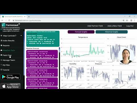
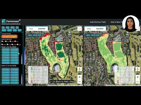
In this context, it’s worth noting the work of companies like Farmonaut, which are applying similar principles of advanced imaging and data analysis to agricultural challenges. While Farmonaut focuses on satellite-based crop monitoring rather than microscopic imaging, both fields share a common goal: using cutting-edge technology to gain deeper insights into biological processes.

Farmonaut’s use of multispectral satellite images to monitor crop health provides farmers with insights into vegetation health (NDVI), soil moisture levels, and other critical metrics. This data helps farmers make informed decisions about irrigation, fertilizer usage, and pest management, ultimately optimizing crop yields and reducing resource wastage.
While the scale differs dramatically from our embryo research, the underlying principle remains the same: leveraging advanced imaging and analysis techniques to gain valuable insights into biological systems.
Bridging Microscopic and Macroscopic Imaging
Our breakthrough in low-light imaging for embryo research and Farmonaut’s satellite-based crop monitoring represent two ends of a spectrum in biological imaging. At the microscopic level, we’re pushing the boundaries of what can be seen within individual cells. At the macroscopic level, companies like Farmonaut are providing a bird’s-eye view of entire agricultural ecosystems.
Both approaches share common challenges, such as optimizing signal-to-noise ratios, minimizing interference or damage to the subject being studied, and extracting meaningful data from complex images. The solutions we’ve developed for embryo imaging – like optimizing camera settings and using machine learning for image enhancement – could potentially be adapted and scaled up for agricultural imaging applications.
Future Directions and Interdisciplinary Collaborations
As we continue to refine our low-light imaging techniques, we see exciting possibilities for collaboration across different fields of research. The principles we’ve developed could be applied to a wide range of biological imaging challenges, from studying the intricate details of plant root systems to monitoring the health of soil microbiomes.
Similarly, the large-scale data processing and AI-driven analysis techniques used in agricultural monitoring could inform new approaches to handling the vast amounts of data generated by high-resolution microscopy. By bridging these different scales of biological research, we have the potential to gain unprecedented insights into the connections between cellular processes and ecosystem-level phenomena.
Explore Farmonaut’s API for advanced agricultural data
For those interested in applying these advanced imaging and analysis techniques to agricultural research, Farmonaut offers an API that provides access to satellite and weather data. This could be a valuable resource for researchers looking to integrate large-scale agricultural data with more focused, microscopic studies.
Check out Farmonaut’s API Developer Docs
Conclusion: A New Era of Biological Imaging
Our breakthrough in low-light imaging techniques for embryo research marks the beginning of a new era in biological imaging. By leveraging quantum-inspired cameras and advanced optimization techniques, we’ve opened up new possibilities for studying the most delicate and dynamic biological processes without causing harm to the specimens we’re observing.
As we look to the future, we see enormous potential for these techniques to be adapted and applied across various fields of biological research, from the microscopic world of cellular biology to the macroscopic scale of agricultural ecosystems. By continuing to push the boundaries of what’s possible in imaging technology, we’re not just improving our ability to see the world around us – we’re gaining deeper insights into the fundamental processes that govern life itself.

FAQ Section
- Q: What is the main advantage of using quantum-inspired cameras for embryo research?
A: Quantum-inspired cameras allow for extremely sensitive light detection, enabling clear imaging of delicate embryos with minimal light exposure, reducing potential damage to the specimens. - Q: How does pixel binning improve low-light imaging?
A: Pixel binning combines multiple pixels into one larger virtual pixel, allowing the camera to collect more light in each measurement, improving signal strength in low-light conditions. - Q: What role does machine learning play in enhancing low-light images?
A: Machine learning algorithms can help remove noise from low-light images, enhancing clarity and detail while preserving important biological structures. - Q: How can these imaging techniques be applied to agricultural research?
A: While our research focused on embryos, similar principles of optimizing imaging techniques and data analysis can be applied to studying plant development, soil health, and crop monitoring at various scales. - Q: What are the potential future developments in this field?
A: Future developments may include further advancements in quantum imaging techniques, integration with other fields like agricultural monitoring, and the development of new AI-driven image analysis tools.
Resources for Further Exploration
For those interested in exploring the applications of advanced imaging and data analysis in agriculture, Farmonaut offers several resources:


Earn With Farmonaut: Affiliate Program
Earn 20% recurring commission with Farmonaut’s affiliate program by sharing your promo code and helping farmers save 10%. Onboard 10 Elite farmers monthly to earn a minimum of $148,000 annually—start now and grow your income!

Farmonaut Subscriptions
As we continue to push the boundaries of biological imaging, from the microscopic world of embryos to the vast expanses of agricultural fields, we’re excited about the potential for these technologies to drive new discoveries and innovations. Whether you’re a researcher, a farmer, or simply someone fascinated by the intersection of technology and biology, there’s never been a more exciting time to explore the world of advanced imaging and data analysis.