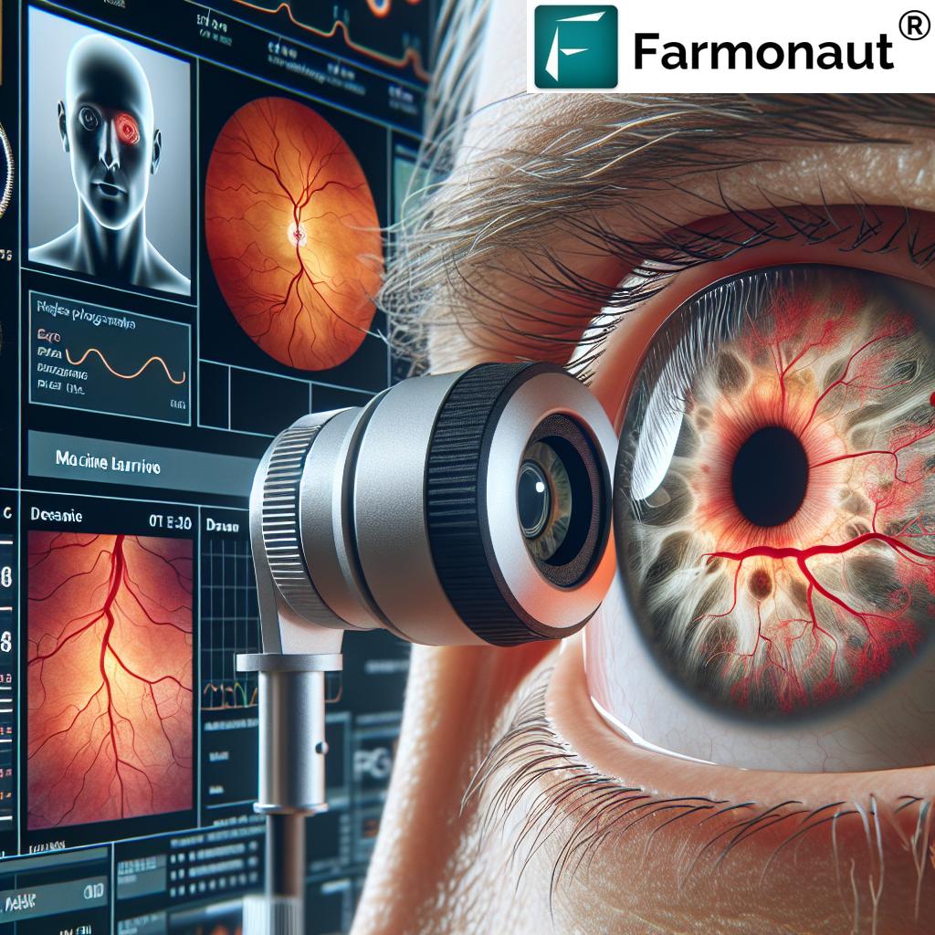
Revolutionizing Stroke Prevention: How Routine Eye Tests in Melbourne Could Save Lives
“Machine learning analysis of 45,000+ retinal images identified 29 indicators linked to first-time stroke risk.”
In the bustling city of Melbourne, Australia, a groundbreaking discovery is set to transform the way we approach stroke prevention. We’re excited to share with you the results of an international study that could revolutionize how we assess stroke risk – and it all starts with a simple eye test.
The Eye-Opening Research
Led by the esteemed Centre for Eye Research Australia (CERA) in Melbourne, this study has unveiled a fascinating connection between our eyes and our risk of stroke. By analyzing what researchers call a unique “fingerprint” of blood vessels at the back of the eye, healthcare professionals can now predict an individual’s stroke risk with an accuracy comparable to traditional risk factors.
What makes this discovery particularly exciting is its non-invasive nature. Unlike some traditional methods that might require blood tests or more complex procedures, this new approach utilizes fundus photography – a standard technique used in routine eye examinations.

The Power of Machine Learning in Eye Exams
At the heart of this research is an innovative tool known as the Retina-based Microvascular Health Assessment System (RMHAS). This sophisticated machine learning system was developed to analyze fundus images with incredible precision.
“The Retina-based Microvascular Health Assessment System discovered 17 indicators related to vascular density for stroke prediction.”
The international research team involved in the study examined data from an impressive 45,161 participants in the UK, with an average age of 55. Over an average follow-up period of 12.5 years, 749 cases of stroke were recorded among the participants.
Unveiling the Retinal Indicators
Out of 118 assessed indicators of vascular health revealed through retinal analysis, the researchers identified 29 that were notably associated with the risk of first-time stroke. Remarkably, about 17 of these indicators were linked to vascular density – the amount of tissue occupied by blood vessels in the retina.
This discovery highlights a crucial connection: a low density of blood vessels, both in the retina and the brain, correlates with a heightened risk of stroke. Each alteration in the vascular density indicators presents an increased stroke risk ranging between 10 to 19 percent.
Beyond Density: Complexity and Twistedness
The research didn’t stop at vascular density. It also revealed that decreases in the complexity and twistedness of these vascular indicators are associated with an increase in stroke risk ranging from 10.5 to 19.5 percent. These correlations underscore the significant role that retinal health plays in overall vascular health and stroke prevention.
A Practical Approach to Stroke Risk Assessment
What makes this model particularly exciting is its practicality. Age and sex are fundamental demographic parameters that are easily obtainable, and retinal parameters can be readily acquired through routine fundus photography. This makes the RMHAS model an incredibly practical approach for stroke risk assessment, especially beneficial for primary healthcare systems and settings with limited resources.
By providing a non-invasive screening method for identifying individuals at risk of stroke, this research opens up new possibilities for early intervention and prevention strategies.
The Global Impact of Strokes
To understand the significance of this research, it’s crucial to consider the global impact of strokes. Strokes affect over 100 million individuals worldwide and result in approximately 6.7 million deaths each year. These alarming statistics underscore the urgent need for effective early identification and prevention strategies.
Given the severity of this health challenge, the potential of routine eye tests to revolutionize stroke prevention cannot be overstated. By utilizing simple eye examinations as a means to detect individuals at risk, healthcare systems could facilitate timely interventions and potentially save millions of lives.
Comparing Traditional and Retinal Indicators
To better understand how this new method compares to traditional risk assessment, let’s take a look at this comparison table:
| Risk Factor Type | Specific Indicator | Relative Risk Score (1-10) | Ease of Detection | Cost-Effectiveness |
|---|---|---|---|---|
| Traditional | Blood Pressure | 8 | High | Medium |
| Traditional | Cholesterol Levels | 7 | Medium | Low |
| Traditional | Smoking Status | 6 | High | High |
| Traditional | Diabetes | 7 | Medium | Medium |
| Traditional | Obesity | 6 | High | High |
| Retinal | Vascular Density | 8 | High | High |
| Retinal | Vessel Complexity | 7 | High | High |
| Retinal | Vessel Twistedness | 7 | High | High |
| Retinal | Microvascular Changes | 8 | High | High |
| Retinal | Retinal Tissue Health | 7 | High | High |
As we can see from this table, retinal indicators offer high ease of detection and cost-effectiveness across the board, while providing comparable risk assessment to traditional factors.
The Future of Stroke Prevention
The implications of this research are far-reaching. By integrating routine eye tests into regular health check-ups, we could potentially identify individuals at risk of stroke much earlier and more efficiently. This early detection could lead to more timely interventions, lifestyle changes, and preventative treatments, ultimately reducing the global burden of stroke.
The Role of Technology in Healthcare
This groundbreaking research showcases the invaluable role that technology plays in advancing healthcare. By leveraging machine learning and digital imaging technologies, we’re able to extract crucial health information from something as simple as an eye test.
It’s worth noting that while this research focuses on stroke prediction, the potential applications of this technology could extend to other areas of health assessment. The retina, often referred to as the “window to the brain,” could provide insights into various neurological and vascular conditions.
Implementing the Findings
While the research results are promising, implementing these findings on a large scale will require coordination between various stakeholders in the healthcare system. Here are some key steps that would be necessary:
- Training healthcare professionals: Optometrists and other eye care professionals would need to be trained in using the RMHAS and interpreting its results.
- Updating guidelines: Health organizations would need to update their guidelines to include retinal screening as part of routine health check-ups, especially for individuals over 50 or those with other risk factors.
- Public awareness: Educating the public about the importance of regular eye exams, not just for vision health but for overall health assessment.
- Technology integration: Ensuring that healthcare facilities have access to the necessary technology and software to perform these advanced analyses.
- Research continuation: Further studies to refine the model and potentially expand its applications to other health conditions.

The Melbourne Connection
It’s particularly exciting that this research has its roots in Melbourne, Australia. The city has long been known for its world-class medical research facilities, and this study further cements its position as a leader in innovative healthcare solutions.
The success of this research could potentially lead to increased funding and support for similar studies in the future, not just in Melbourne but around the world. It also highlights the importance of international collaboration in medical research, as this study involved data from the UK and expertise from various countries.
Beyond Stroke: Potential Applications
While the focus of this research has been on stroke prevention, the implications could be much broader. The retina’s unique position as a window into our vascular health means that this type of screening could potentially be used to assess risk for other conditions as well. Some areas that might benefit from similar research include:
- Cardiovascular disease: Many of the risk factors for stroke are also risk factors for heart disease.
- Diabetes: Retinal changes are already used to diagnose diabetic retinopathy, but this more detailed analysis could provide earlier warnings.
- Hypertension: Changes in retinal blood vessels can be an early indicator of high blood pressure.
- Cognitive decline: Some studies have suggested links between retinal changes and conditions like Alzheimer’s disease.
The Importance of Regular Eye Tests
This research underscores the importance of regular eye tests, not just for maintaining good vision, but as a crucial part of overall health monitoring. Here are some key reasons why everyone should prioritize routine eye exams:
- Early detection of eye diseases: Many eye conditions, such as glaucoma and macular degeneration, can be treated more effectively if caught early.
- Updating prescriptions: Regular check-ups ensure that your glasses or contact lens prescriptions are up to date, preventing eye strain and headaches.
- Detecting systemic health issues: As this research shows, eye exams can reveal signs of broader health problems, including diabetes, high blood pressure, and now, stroke risk.
- Preventive care: Regular exams allow for preventive measures to be taken before vision problems develop or worsen.
- Digital eye strain assessment: With increasing screen time, optometrists can provide advice on managing digital eye strain.
The Role of AI and Machine Learning in Healthcare
The RMHAS system used in this study is a prime example of how artificial intelligence (AI) and machine learning are revolutionizing healthcare. These technologies are enabling us to:
- Analyze vast amounts of data: Machine learning can process and find patterns in datasets far larger than humans could manually analyze.
- Identify subtle indicators: AI can detect minute changes or patterns that might be missed by human observers.
- Predict outcomes: By learning from large datasets, AI can make predictions about future health outcomes.
- Personalize treatment: AI can help tailor treatments to individual patients based on their unique characteristics.
- Improve efficiency: Automated systems can speed up analysis and free up healthcare professionals to focus on patient care.
Challenges and Considerations
While the potential of this research is enormous, it’s important to consider some of the challenges that might arise in implementing this technology on a large scale:
- Data privacy: As with any health data, ensuring the privacy and security of retinal scans will be crucial.
- Ethical considerations: There may be ethical questions about how this information is used, particularly in contexts like insurance or employment.
- Access to technology: Ensuring that this technology is available in all healthcare settings, including rural and low-resource areas, will be a challenge.
- Integration with existing systems: The new screening method will need to be integrated smoothly with existing healthcare protocols and electronic health record systems.
- False positives and negatives: As with any screening tool, there will be a need to manage the risk of false positives (which could cause unnecessary anxiety) and false negatives (which could provide false reassurance).
The Global Impact
The potential global impact of this research cannot be overstated. Stroke is a leading cause of death and disability worldwide, with a particularly heavy burden in low- and middle-income countries. A cost-effective, non-invasive screening method could be a game-changer in these settings.
Moreover, as populations age globally, the incidence of stroke is likely to increase. This makes preventive strategies even more crucial. By identifying at-risk individuals early, healthcare systems could potentially reduce the burden of stroke care and rehabilitation, leading to significant cost savings and improved quality of life for millions of people.
Next Steps in Research
While this study represents a significant breakthrough, it’s likely just the beginning of a new frontier in stroke prevention and vascular health assessment. Some potential next steps in this field of research might include:
- Longitudinal studies: Following participants over even longer periods to further validate the predictive power of retinal indicators.
- Intervention studies: Researching whether early interventions based on retinal screening can indeed reduce stroke incidence.
- Expanding the model: Incorporating additional data points to potentially improve the accuracy of risk prediction.
- Cross-cultural studies: Validating the model across different populations and ethnic groups.
- Technology development: Creating more accessible and affordable retinal imaging technologies to facilitate widespread adoption.
FAQ Section
Q: How accurate is this new method of stroke risk assessment?
A: The study shows that retinal indicators can predict stroke risk with accuracy comparable to traditional risk factors.
Q: Do I need a special eye exam for this screening?
A: No, the screening uses fundus photography, which is commonly employed in standard eye examinations.
Q: At what age should I start getting these screenings?
A: While the study focused on individuals with an average age of 55, it’s always best to consult with your healthcare provider about when to start screenings based on your individual risk factors.
Q: Can this method detect other health issues besides stroke risk?
A: While this study focused on stroke risk, retinal exams can potentially provide insights into other vascular and neurological conditions. Further research is ongoing.
Q: How often should I get my eyes checked for this purpose?
A: The frequency of eye exams can vary based on age and risk factors. Generally, adults should have a comprehensive eye exam every 1-2 years, but consult with your eye care professional for personalized advice.
Conclusion
The groundbreaking research from Melbourne’s Centre for Eye Research Australia represents a significant leap forward in stroke prevention. By leveraging the power of routine eye tests and advanced machine learning, we now have a promising new tool in the fight against one of the world’s leading causes of death and disability.
This innovative approach not only offers a non-invasive and cost-effective method of assessing stroke risk but also opens up new avenues for early intervention and prevention. As we move forward, the integration of this technology into routine healthcare could potentially save countless lives and significantly reduce the global burden of stroke.
The future of healthcare is here, and it’s clear that our eyes hold more secrets to our overall health than we ever imagined. Regular eye tests are no longer just about maintaining good vision – they could be the key to unlocking a healthier, longer life.
As we continue to explore the potential of this groundbreaking research, one thing is certain: the way we approach stroke prevention and overall health assessment is set to change dramatically. And it all starts with a simple look into our eyes.


